Seeing beyond BMI: Estimating cardiometabolic risk with smartphone imagery
Smartphone imagery may offer a more nuanced view of cardiometabolic risk than BMI alone. This article examines the promise, safety considerations, and limitations of using AI-driven visual assessment in everyday health monitoring.
Tags
Quick summary
Smartphone imagery may offer a more nuanced view of cardiometabolic risk than BMI alone. This article examines the promise, safety considerations, and limitations of using AI-driven visual assessment in everyday health monitoring.
Seeing beyond BMI: Estimating cardiometabolic risk with smartphone imagery
For decades, body mass index (BMI) has been the default shorthand for metabolic health. Clinicians calculate it in seconds, insurers use it to adjust premiums, and public health agencies rely on it to map obesity trends. Yet BMI has always been a blunt instrument. It cannot distinguish muscle from fat, say nothing about where fat is distributed, and often misclassifies individuals who are metabolically healthy or unhealthy. The search for a better, more accessible proxy has been ongoing for years. A particularly intriguing candidate has now emerged from the intersection of machine learning and everyday technology: the smartphone camera. In August 2026, Google Research published a blog post titled "Seeing beyond BMI: Estimating cardiometabolic risk with smartphone imagery," describing how ordinary photographs taken with a mobile device could potentially provide a far richer signal about cardiometabolic risk than a simple height-and-weight calculation ever could.
The idea is not merely to refine an old metric, but to rethink what kind of data we can extract from the human body using nothing more than a camera. This article explores why BMI falls short, how smartphone imagery could fill the gap, and what must happen before such technology moves from the research blog to the clinic.
Why BMI falls short as a health marker
BMI was introduced in the nineteenth century by the Belgian statistician Adolphe Quetelet, who was interested in defining the "normal" human physique across populations. The formula—weight in kilograms divided by height in meters squared—was never designed to diagnose disease. It was a population-level statistical tool, not a personalized medical assessment. That distinction matters now more than ever.
Two individuals with identical BMIs can have wildly different health profiles. A rugby player and a sedentary office worker might share the same height and weight, yet the former carries substantial muscle mass while the latter carries excess visceral fat. Their risks for type 2 diabetes, hypertension, and cardiovascular disease diverge sharply, but BMI alone cannot capture that difference. Likewise, people with "normal" BMIs can still have metabolic syndrome if they carry fat centrally—a condition sometimes called normal-weight obesity. Studies consistently show that waist circumference and waist-to-hip ratio correlate more strongly with adverse cardiometabolic outcomes than BMI does. The reason is anatomical: visceral fat surrounding the liver, pancreas, and intestines is metabolically active, secreting inflammatory cytokines and free fatty acids that impair insulin sensitivity. Subcutaneous fat, by contrast, is comparatively benign.
This has led researchers to search for better proxies. DEXA scans, MRI, and CT imaging can precisely measure body composition, but they are expensive, require specialized equipment, and cannot easily scale to large populations. Bioelectrical impedance scales are cheaper but notoriously finicky, varying with hydration and electrode placement. Tape measures for waist circumference are simple but prone to user error. What the field really needs is a method that is accurate, scalable, and accessible to billions of people worldwide. Smartphone imagery, powered by modern computer vision, may be the first candidate that satisfies all three criteria.
Smartphone imagery: a new window on body composition
The core premise of the Google Research initiative is deceptively simple: the human body carries visible, externally observable clues about internal cardiometabolic health. Fat distributes in patterns—around the abdomen, the hips, the thighs—that are not random. Muscle mass shapes contours. Posture reflects both skeletal alignment and soft tissue composition. These cues have always been readable by a trained eye, but they are subtle and difficult to quantify with a tape measure. A camera, however, can capture them all in a single frame.
Modern computer vision models excel at precisely this kind of task. Given a large dataset of images paired with known health outcomes, a neural network can learn to associate specific visual features with underlying physiological states. The network does not need an explicit set of rules about where the waist ends and the hip begins. Instead, it learns from patterns in millions of examples. The result is a model that can infer body composition and cardiometabolic risk factors from images alone, with no physical contact, no specialized hardware, and no clinical training required by the operator.
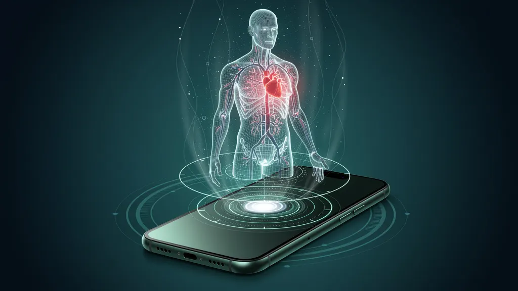
The practical implications are substantial. A person could take a standard smartphone photograph of their torso, from the front or the side, and receive an estimate of body fat percentage, waist-to-hip ratio, or even a composite cardiometabolic risk score—all without stepping into a clinic. This is not an unrealistic fantasy. The underlying technology—convolutional neural networks, pose estimation, and image segmentation—has matured dramatically over the past decade. Models can now identify anatomical landmarks, segment body regions, and estimate joint positions with remarkable accuracy. Extending that capability to estimate soft tissue composition is a natural next step.
From pixels to risk profiles: what the machine learns
One of the most compelling aspects of this approach is that the model may identify visual patterns that human clinicians rarely consider explicitly. BMI focuses on overall mass, but a person's silhouette encodes far more information. The ratio between shoulder width and waist width, the curvature of the abdomen, the thickness of the neck, the accumulation of fat around the flanks—these are all visually available in a single photograph. A machine learning model can weigh dozens of such features simultaneously and produce a risk estimate that integrates more information than any human eye could deliberately evaluate.
This is analogous to how deep learning has transformed medical imaging in other domains. Radiologists once relied on their trained eyes to spot tumors in X-rays and CT scans. Today, algorithms can detect subtle patterns in those same images that human radiologists miss, sometimes flagging early-stage disease that would otherwise go unnoticed. The same principle applies to body imagery. The visual features predictive of insulin resistance or fatty liver disease may be subliminal—not obvious to a physician, but consistent enough for a well-trained model to latch onto them.
Importantly, the model can output continuous scores rather than categorical labels. Instead of saying a person falls into "overweight" or "obese" based on an arbitrary cutoff, the system might produce a risk percentile or a calibrated probability of future cardiometabolic events. This aligns with the broader shift in medicine toward personalized, continuous risk stratification rather than binary thresholds. It also allows the model to be tuned for different populations. A visual risk model trained primarily on images from one ethnic group may perform poorly on another, but with sufficient and diverse training data, the model can learn population-specific patterns.
Why this matters for global health
The greatest promise of smartphone-based risk estimation lies in its accessibility. More than six billion people worldwide own smartphones, and the number continues to grow. Many of these people live in regions where access to clinical diagnostic tools is limited. An MRI machine costs millions of dollars and requires infrastructure, trained technicians, and a stable power supply. A smartphone is cheap, ubiquitous, and carried everywhere. If cardiometabolic risk can be estimated from a simple photograph, then screening could be democratized in ways that were previously unimaginable.
Consider a rural health worker in a low-income country who has been trained to spot malnutrition but lacks any diagnostic equipment. With a smartphone-based risk assessment tool, that worker could screen entire villages for cardiometabolic risk within hours. The data could be aggregated anonymized, providing public health authorities with unprecedented visibility into the metabolic health of their populations. In high-income countries, the same technology could enable continuous self-monitoring, allowing individuals to track changes in their risk profile over time and to see the effects of lifestyle interventions in near real time.
The Google Research blog post represents an important step in this direction. It positions the work not merely as an interesting computer vision exercise, but as a serious contribution to preventive medicine. The underlying message is that the tools for transforming global health may already be sitting in our pockets. What has been missing is the algorithmic insight to unlock their diagnostic potential.
Privacy, bias, and ethical considerations
No discussion of health-related machine learning is complete without addressing the ethical and societal risks. Smartphone imagery of the human body is inherently sensitive. Body images are deeply personal, and a tool that asks users to photograph themselves inevitably raises privacy concerns. Who has access to those images? Where are they stored? Can they be deleted? Can they be re-purposed for advertising or insurance underwriting? These are not hypothetical questions; they are the kind of concerns that can derail a promising technology if not addressed thoughtfully.
There is also the risk of algorithmic bias. A model trained on images from a certain demographic—say, predominantly lighter-skinned, North American bodies—may perform poorly on dark-skinned individuals or on bodies with different average proportions. Machine learning systems are notorious for inheriting the biases of their training data. If a smartphone-based risk assessment tool systematically underestimates risk for certain populations, it could exacerbate existing health disparities rather than reduce them. Researchers must therefore be meticulous about collection of diverse training data and about evaluating model performance across demographic subgroups.
A further concern is medical and regulatory oversight. If a tool provides estimates of cardiometabolic risk, it is effectively a medical device, whether or not it is labeled as such. Regulators in the United States, the European Union, and elsewhere have developed pathways for software as a medical device, but these pathways are still evolving. A smartphone app that gives risk scores might require clinical validation and regulatory approval before it can be marketed to consumers. That approval process is slow and costly, but it is essential for protecting public safety.
From research to clinical reality
It is worth remembering that the Google Research blog post describes research in progress, not a product that is available today. Many technical and practical hurdles remain between a promising prototype and a widely deployed clinical tool. The accuracy of image-based risk estimation must be rigorously validated against gold-standard cardiometabolic measurements—blood panels, DEXA scans, and longitudinal health outcomes. The models must be compressed to run efficiently on-device, so that users do not need to upload sensitive images to the cloud. And the user experience must be designed to be foolproof, because the quality of the input image—lighting, angle, distance, clothing—will directly affect the quality of the output estimate.
None of these challenges are insurmountable, but they require time, funding, and cross-disciplinary collaboration between machine learning researchers, physicians, epidemiologists, and privacy advocates. The trajectory is nevertheless promising. The broader pattern in medicine is unmistakable: artificial intelligence is moving from the laboratory into everyday clinical and consumer contexts at an accelerating pace. Smartphone cardiometabolic risk estimation fits squarely within that trend.
Perhaps the most important contribution of the Google Research work is conceptual. By shifting the frame from BMI to image-based risk assessment, the researchers are challenging the field to think more ambitiously about what constitutes health data. A photograph is a form of vitals—a compressed record of physiological state that can be unpacked by the right algorithm. If that vision is realized, the humble act of taking a selfie could one day yield insights that a medical chart cannot.
Conclusion
BMI has served public health for over a century, but its limitations are increasingly difficult to ignore in an era of personalized medicine and affordable computing. The Google Research initiative described in "Seeing beyond BMI: Estimating cardiometabolic risk with smartphone imagery" offers a powerful alternative. By applying modern computer vision to ordinary photographs, it is possible to move beyond crude weight-based categories and toward nuanced, continuous estimates of cardiometabolic risk.
The technology is not ready for prime time. It must still overcome significant obstacles around validation, privacy, bias, and regulation. But the direction is clear, and the potential benefits are enormous. In a world where cardiovascular disease remains the leading cause of death and diabetes affects hundreds of millions of people, any tool that can make screening faster, cheaper, and more accessible deserves close attention. A camera phone in every pocket may not be the first thing that comes to mind when we think of medical diagnostics. But if this research bears fruit, it could become the most ubiquitous health monitoring device ever created.